The human skull is a complex structure that serves multiple critical functions, including protect the brain and providing support for facial structures. Among the various components of the skull, the bones of the calvaria play a polar role in safeguarding the brain. The calvaria, also known as the cranial vault, consists of eight bones that form the roof and sides of the skull. Understanding the anatomy, functions, and clinical signification of these bones is indispensable for medical professionals and students alike.
The Anatomy of the Bones of the Calvaria
The calvaria is indite of eight bones, which are couple except for the frontal and occipital bones. These bones are:
- Frontal Bone: Forms the forehead and the roof of the orbits (eye sockets).
- Parietal Bones (2): Form the sides and roof of the skull.
- Temporal Bones (2): Located on the sides of the skull, below the parietal bones.
- Occipital Bone: Forms the back and establish of the skull.
- Sphenoid Bone: Located at the free-base of the skull, behind the eyes.
- Ethmoid Bone: Located between the eyes, forming part of the adenoidal cavity and the roof of the orbits.
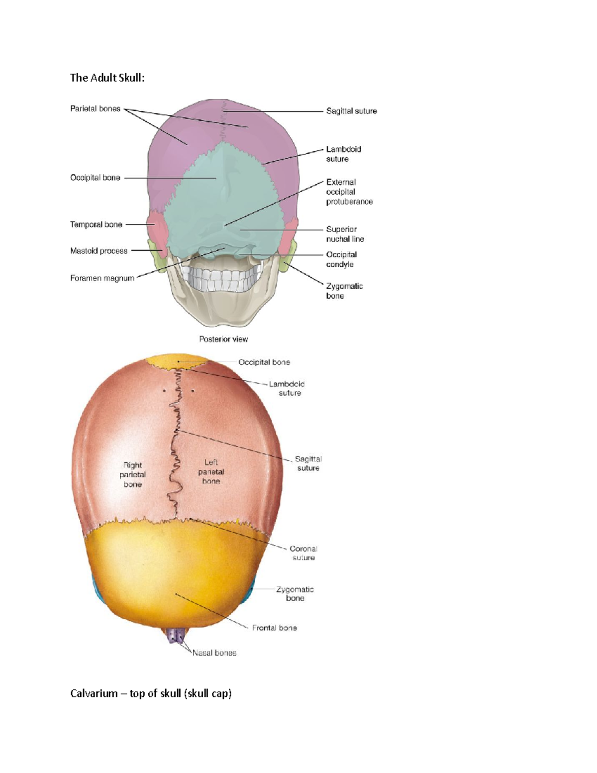
The bones of the calvaria are associate by sutures, which are unchewable joints that let for some movement during growth and development. The major sutures of the calvaria include:
- Coronal Suture: Between the head-on and parietal bones.
- Sagittal Suture: Between the two parietal bones.
- Lambdoid Suture: Between the parietal and occipital bones.
- Squamous Suture: Between the parietal and temporal bones.
Functions of the Bones of the Calvaria
The main office of the bones of the calvaria is to protect the brain from external impacts and injuries. Additionally, these bones provide attachment sites for muscles and ligaments that back the head and neck. The calvaria also plays a role in the production of red blood cells, as the marrow within the bones can make blood cells during foetal development and betimes childhood.
Another significant function of the calvaria is its role in the growth and development of the skull. The sutures between the bones allow for the expansion of the skull as the brain grows, ensuring that the brain has adequate space to develop decent.
Clinical Significance of the Bones of the Calvaria
The bones of the calvaria are crucial in diverse clinical contexts, including trauma, surgery, and developmental disorders. Understanding the anatomy and functions of these bones is essential for diagnosing and treat conditions that regard the skull.
Trauma: Injuries to the calvaria can result in fractures, which may be linear, depressed, or grind. These fractures can cause significant damage to the brain and require immediate medical attention. Common causes of calvarial fractures include falls, motor vehicle accidents, and assaults.
Surgery: Surgical procedures involving the calvaria are often perform to access the brain or to repair fractures. Craniotomy, for instance, is a surgical procedure in which a portion of the calvaria is temporarily withdraw to access the brain. This procedure is ordinarily used to treat brain tumors, aneurysms, and other neurologic conditions.
Developmental Disorders: Abnormalities in the development of the bones of the calvaria can lead to various congenital disorders. for case, craniosynostosis is a condition in which one or more of the sutures fuse untimely, leading to unnatural skull shape and likely brain development issues. Early diagnosis and treatment of craniosynostosis are crucial to prevent complications.
Diagnostic Imaging of the Bones of the Calvaria
Diagnostic visualize plays a crucial role in appraise the bones of the calvaria. Various imaging modalities are used to assess the construction and unity of these bones, include:
- X rays: Provide a canonic overview of the skull's bony structures and can detect fractures and other abnormalities.
- Computed Tomography (CT) Scans: Offer detail images of the skull and can detect subtle fractures, bone lesions, and other abnormalities.
- Magnetic Resonance Imaging (MRI): Provides detailed images of the brain and border structures, helping to assess the extent of injuries and detect soft tissue abnormalities.
Imaging studies are crucial for name and manage conditions affect the bones of the calvaria. They assist aesculapian professionals determine the earmark course of treatment and reminder the progress of heal or disease.
Common Conditions Affecting the Bones of the Calvaria
Several conditions can affect the bones of the calvaria, ranging from traumatic injuries to developmental disorders. Some of the most common conditions include:
- Fractures: Can occur due to trauma and may be linear, depressed, or mash.
- Craniosynostosis: Premature fusion of the sutures, leading to abnormal skull shape and possible brain development issues.
- Osteomyelitis: Infection of the bone, which can make pain, swell, and fever.
- Metastatic Cancer: Cancer that has spread from another part of the body to the bones of the calvaria.
- Paget's Disease: A inveterate condition that causes abnormal bone growth and can regard the bones of the calvaria.
Each of these conditions requires specific diagnostic and treatment approaches. Early detection and intervention are crucial for managing these conditions effectively.
Treatment Options for Conditions Affecting the Bones of the Calvaria
Treatment options for conditions affecting the bones of the calvaria vary bet on the underlie cause and severity of the condition. Some common treatment approaches include:
- Surgical Intervention: May be postulate for fractures, craniosynostosis, and other structural abnormalities. Surgical procedures can regard repair fractures, removing bone fragments, or reshape the skull.
- Medications: Antibiotics may be prescribed for infections such as osteomyelitis. Pain management medications may also be used to assuage discomfort.
- Radiation Therapy: May be used to treat metastatic crab that has spread to the bones of the calvaria.
- Physical Therapy: Can facilitate better mobility and strength after injuries or surgeries.
Treatment plans are tailored to the individual needs of the patient and may imply a multidisciplinary approach, include input from neurologists, neurosurgeons, orthopedic surgeons, and other specialists.
Prevention and Management of Injuries to the Bones of the Calvaria
Preventing injuries to the bones of the calvaria involves taking precautions to avoid head trauma. Some prophylactic measures include:
- Wearing helmets during activities that pose a risk of head injury, such as cycle, skiing, and contact sports.
- Using seatbelts and child safety seats in vehicles.
- Installing safety features in the home, such as stair gates and non slip mats.
- Avoiding activities that increase the risk of falls, especially for older adults.
If an injury to the bones of the calvaria occurs, prompt aesculapian attending is all-important. Early diagnosis and treatment can prevent complications and improve outcomes. Regular postdate up with healthcare providers is also significant to admonisher mend and detect any possible issues betimes.
Note: Always consult with a healthcare professional for advice tailored to your specific position.
Research and Future Directions
Ongoing research is focalize on ameliorate our understanding of the bones of the calvaria and germinate new treatments for conditions affecting these bones. Advances in imaging engineering, surgical techniques, and medical therapies are paving the way for better diagnosis and management of calvarial disorders.
Future research may explore the use of stem cells and regenerative medicine to repair damage bones and promote cure. Additionally, genetic studies may ply insights into the underlie causes of developmental disorders affecting the bones of the calvaria, leading to new preventive and remedial strategies.
Collaboration between researchers, clinicians, and patients is essential for progress our noesis and improving outcomes for individuals with conditions impact the bones of the calvaria.
Researchers are also investigating the role of the calvaria in brain development and function. Understanding how the skull interacts with the brain can provide valuable insights into neurological disorders and potential treatments.
Advances in 3D printing technology are also being research for the creation of custom implants and prosthetics for patients with complex calvarial injuries or deformities. This engineering has the likely to revolutionise the treatment of calvarial conditions by providing personalise solutions sew to each patient's unequaled anatomy.
besides technological advancements, there is a growing emphasis on preventive measures and public awareness campaigns to cut the incidence of head injuries. Education and outreach programs can assist individuals understand the importance of protect the bones of the calvaria and lead steps to prevent injuries.
Research into the familial and environmental factors that contribute to calvarial disorders is also ongoing. Identifying these factors can aid in the development of target therapies and preventive strategies. for illustration, familial testing may be used to identify individuals at risk for craniosynostosis, countenance for betimes intercession and management.
Collaborative efforts between healthcare providers, researchers, and patients are crucial for progress our understanding of the bones of the calvaria and improving patient outcomes. By working together, we can develop innovative solutions and cater punter care for individuals with calvarial conditions.
Future enquiry may also focus on the long term effects of calvarial injuries and disorders on brain part and overall health. Understanding these effects can assist in the development of comprehensive treatment plans that address both immediate and long term needs.
to resume, the bones of the calvaria play a critical role in protecting the brain and indorse the skull s construction. Understanding their anatomy, functions, and clinical import is essential for name and process conditions that touch these bones. Advances in enquiry and engineering are paving the way for improved diagnosis, treatment, and bar of calvarial disorders, ultimately enhancing the quality of life for individuals touch by these conditions.
Related Terms:
- calvarial anatomy
- where is the calvaria
- bones of calvarium
- right calvarium location
- calvaria sutures
- calvarium of the skull